

KFUPM
KFUPM-SDAIA Joint Research Centre for AI
BRAIN Lab
IEEE JBHI · 2026

PULSE

A Unified Multi-Task Architecture for Cardiac Segmentation, Diagnosis, and Few-Shot Cross-Modality Clinical Adaptation

Hania Ghouse1   Maryam Alsharqi2   Farhad Nezami2,3   Muzammil Behzad1,4,*

1KFUPM, Saudi Arabia  •  2Institute for Medical Engineering and Science, MIT, USA  •  3Harvard Medical School, Harvard University, USA  •  4KFUPM-SDAIA Joint Research Centre for AI
*Corresponding: muzammil.behzad@kfupm.edu.sa

Right ventricle Myocardium Left ventricle

MIT
Harvard
88.8%
ACDC mean Dice (5-fold ensemble)
90.0%
Cardiomyopathy diagnosis accuracy
85.3%
Zero-shot Dice on M&Ms-2 (360)
73.2%
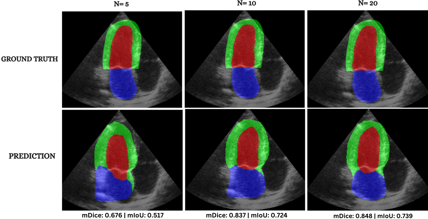
Few-shot Dice on CAMUS (N=20)
Abstract

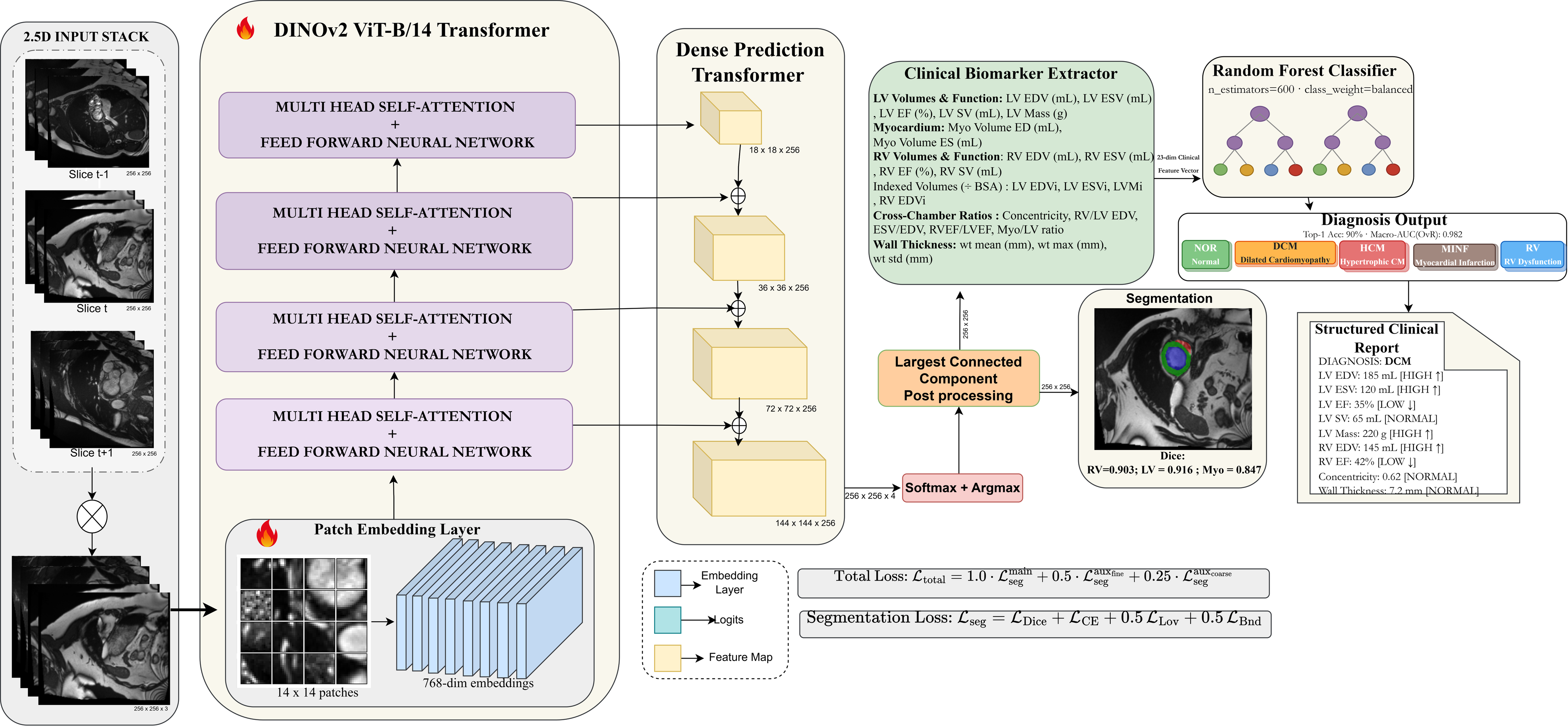
Cardiac image analysis requires accurate ventricular segmentation, disease classification, and structured clinical reporting; these tasks are typically handled by separate models, limiting clinical deployment. To address this, we introduce PULSE, a unified three-task framework that performs: (1) ventricular segmentation using a DINOv2 Vision Transformer (ViT-B/14) backbone with a Dense Prediction Transformer (DPT) decoder and deep supervision; (2) cardiomyopathy diagnosis via a 23-dimensional clinical biomarker vector fed to a Random Forest classifier; and (3) a structured, template-based clinical reporting module that populates a predefined report with the measured indices and rule-based abnormality flags. The framework is entirely vision-based: all reported text is produced by deterministic, rule-driven templates. Additionally, the model takes 2.5D inputs (three adjacent short-axis slices) and is evaluated via a 5-fold stratified ensemble. With extensive experiments on the ACDC benchmark, PULSE achieves a mean Dice of 88.8% (RV: 90.3%, Myo: 84.7%, LV: 91.6%, HD95 ≤ 4.6 mm), 90.0% patient-level diagnostic accuracy (macro-AUC 0.982), and 92.7% clinical flag agreement in the generated reports (LVEF MAE 3.09%, within inter-observer tolerance). Without retraining, PULSE achieves 85.3% mean Dice on M&Ms-2 (360 subjects, RV: 87.9%, Myo: 80.4%, LV: 87.5%) and 88.1% LV Dice on Sunnybrook MRI. We further demonstrate that few-shot fine-tuning on CAMUS echocardiography samples yields a mean Dice of 73.2%, showing strong cross-modality transfer from cardiac MRI priors.

Architecture

One examination in, three clinical outputs out

Framework overview PULSE architecture
A DINOv2 ViT-B/14 encoder with a DPT decoder segments the ventricles from a 2.5D input; clinical biomarkers computed from the masks drive the Random Forest diagnosis and the rule-based clinical report.
Pipeline

Segment, diagnose, report

1. Segment

DINOv2 + DPT delineate RV, myocardium, and LV from three adjacent short-axis slices.

2. Diagnose

A 23-dimensional biomarker vector feeds a Random Forest for patient-level cardiomyopathy diagnosis.

3. Report

Measured indices and rule-based flags populate a structured clinical report, with no language model.

Highlights

What makes PULSE different

3 in 1

Unified multi-task

Segmentation, diagnosis, and reporting from a single model, with no task-specific retraining.

ViT-B/14

Foundation backbone

Self-supervised DINOv2 features, fine-tuned with a two-stage curriculum and a DPT decoder.

23-D

Interpretable diagnosis

Every classifier input is a named clinical measurement: EF, volumes, mass, wall thickness.

5-shot

Cross-modality transfer

Adapts from MRI to echocardiography with only a handful of labels.

Results

Accurate, generalizable, and transferable

ACDC segmentation, 5-fold ensemble with test-time augmentation (50 test patients)
StructureDice (%)IoU (%)HD95 (mm)ASSD (mm)
Left ventricle (LV)91.684.53.870.95
Myocardium (Myo)84.773.53.720.87
Right ventricle (RV)90.382.34.571.05
Mean88.880.14.050.96
Comparison with published methods on ACDC (Dice %)
MethodRVMyoLVMeanDiagnosis
U-Net82.378.794.985.3No
nnU-Net90.188.495.791.4No
TransUNet84.577.794.185.4No
SwinUNet90.779.193.987.9No
PULSE (ours)90.384.791.688.890.0%
Zero-shot generalization (no retraining)
DatasetSubjectsMean Dice
M&Ms-2 (4 vendors)36085.3
Sunnybrook (LV)4588.1
Few-shot CAMUS echocardiography (2-class)
Labelled NMyoLVMean
565.272.168.7
1067.873.370.6
2070.875.673.2
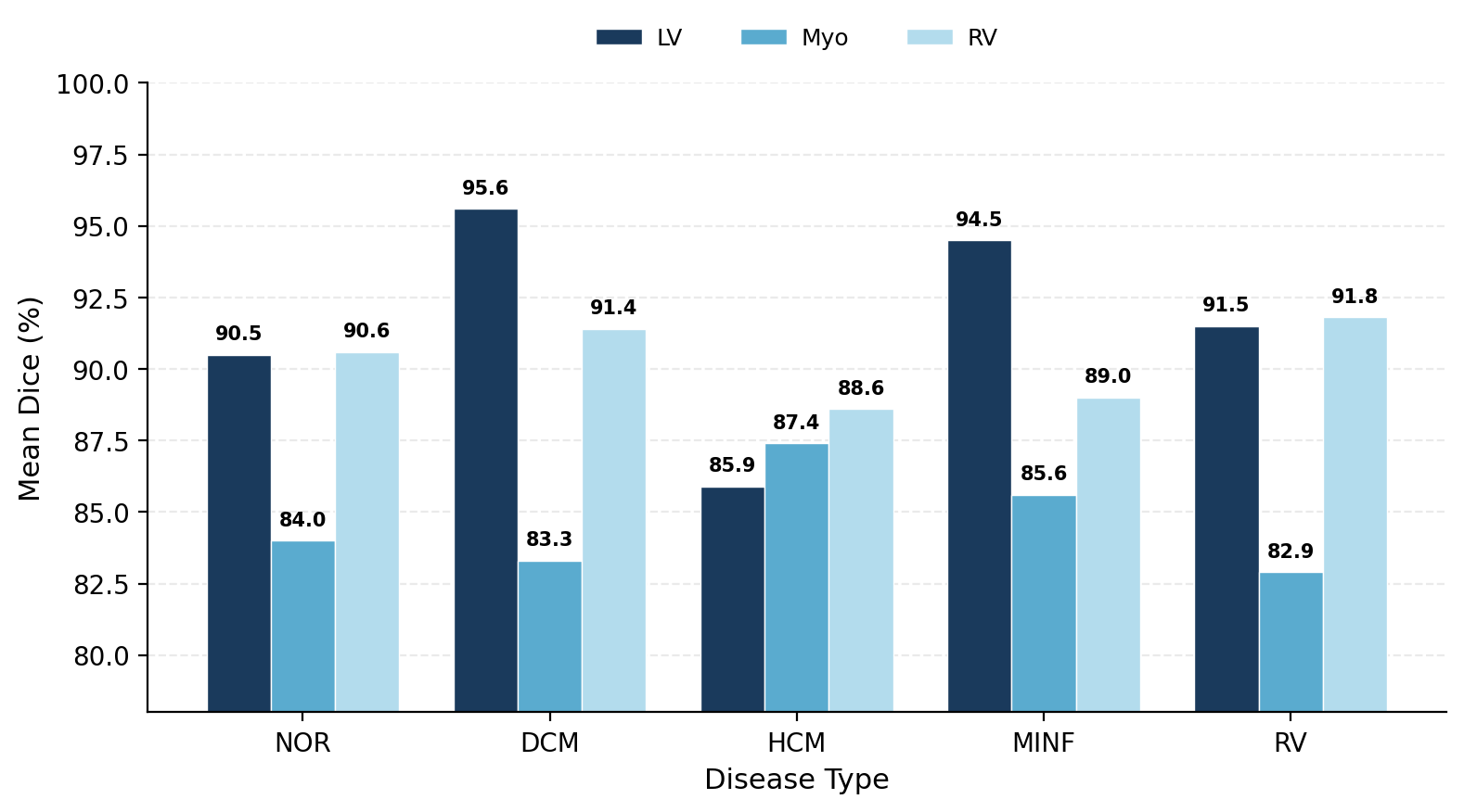
SegmentationPer-disease Dice
Per-disease Dice across the five ACDC classes.
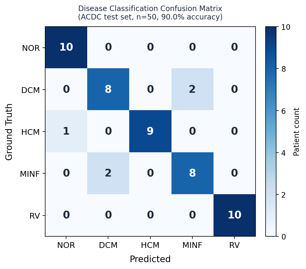
DiagnosisConfusion matrix
Diagnosis confusion matrix on the ACDC test set.
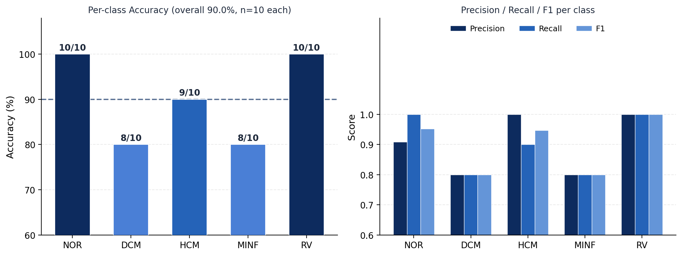
DiagnosisDiagnosis performance
Per-class accuracy, precision, recall, and F1 for cardiomyopathy classification.
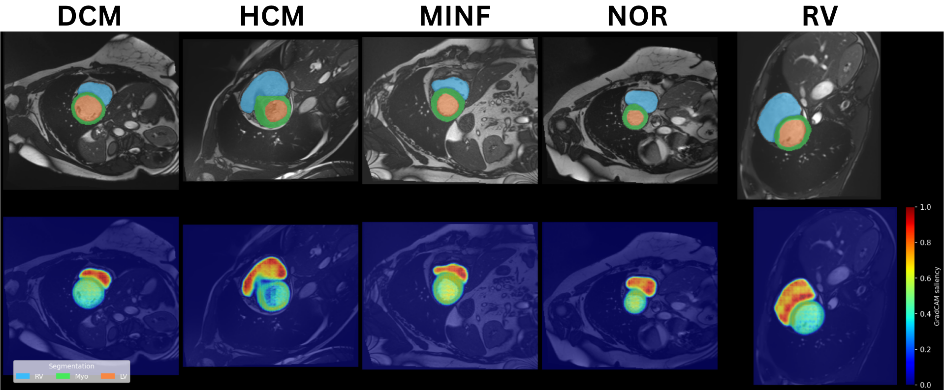
InterpretabilityDINOv2 attention maps
DINOv2 self-attention concentrates on the cardiac region across all disease classes.
Qualitative

Segmentation across vendors and modalities

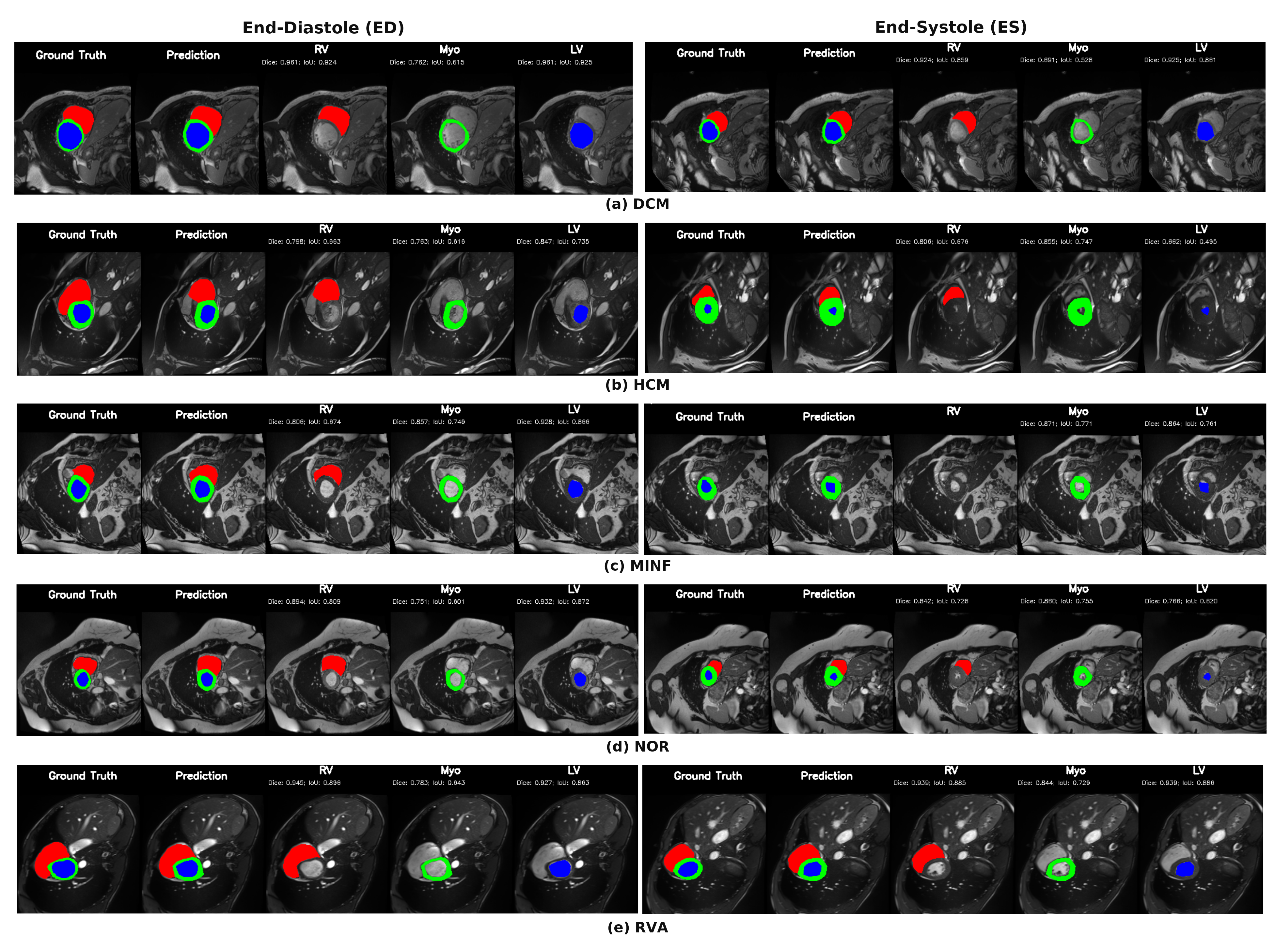
ACDCACDC segmentation across cardiomyopathy classes
End-diastole (ED) and end-systole (ES) segmentation across the five ACDC cardiomyopathy classes (DCM, HCM, MINF, NOR, RVA): RV (red), Myocardium (green), LV (blue).
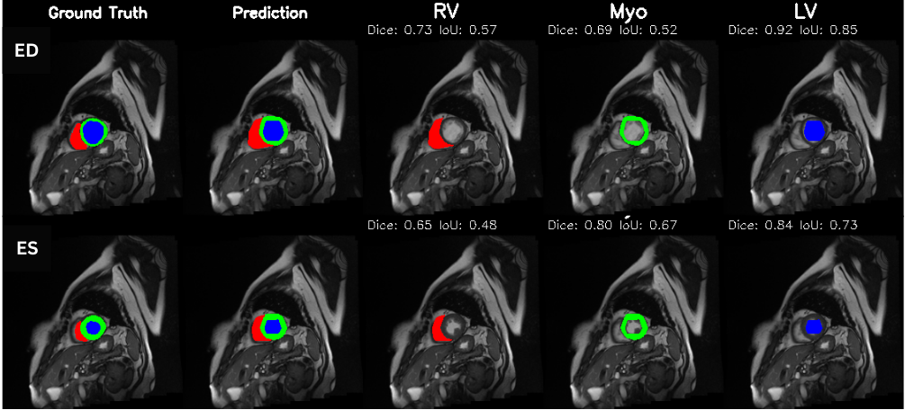
Zero-shot MRIM&Ms-2 generalization
Zero-shot generalization to multi-vendor M&Ms-2 cine MRI: RV (red), Myocardium (green), LV (blue).
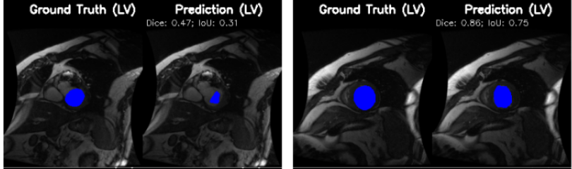
Zero-shot MRISunnybrook segmentation
Zero-shot transfer to the Sunnybrook Cardiac Data with well preserved LV structure.
Few-shot ultrasoundCAMUS few-shot
Few-shot adaptation to CAMUS echocardiography from a handful of labelled scans.
Get started

Code, weights, and data

Citation

Cite PULSE

@article{ghouse2026pulse,
  title   = {PULSE: A Unified Multi-Task Architecture for Cardiac
             Segmentation, Diagnosis, and Few-Shot Cross-Modality
             Clinical Adaptation},
  author  = {Ghouse, Hania and Alsharqi, Maryam and
             Nezami, Farhad and Behzad, Muzammil},
  journal = {IEEE Journal of Biomedical and Health Informatics (JBHI)},
  year    = {2026}
}